Hi!
My name is Emma Linnér and I am currently on my final year of a bachelor in Biology. I am planning to do my masters in neuroscience/cell biology and hoping to pursue a path in academia after that, hopefully getting to work with research. To get an insight in the work life of the people doing research I decided to take the class in research practice BIO299. I was lucky to find a research group working with regeneration of the nervous system and was more than happy when I found out that they had a project suitable for this course and a supervisor that could guide me through the project throughout this autumn. My project was part of a bigger research project studying regeneration of the nervous system in the starlet sea anemone Nematostella vectensis. The purpose of my project was to ablate the nervous system in a group of animals to later observe the regeneration of neurons using a confocal microscope.
Humans, along with many other animals have a relatively poor ability to regenerate lost or damaged tissue. This is not the case for all organisms and some animals possess extraordinary abilities to regenerate tissues after injury or ablation. The starlet sea anemone Nematostella
vectensis has excellent regenerative abilities and can regenerate a whole body axis in less than a week and their nervous system within 25 days.
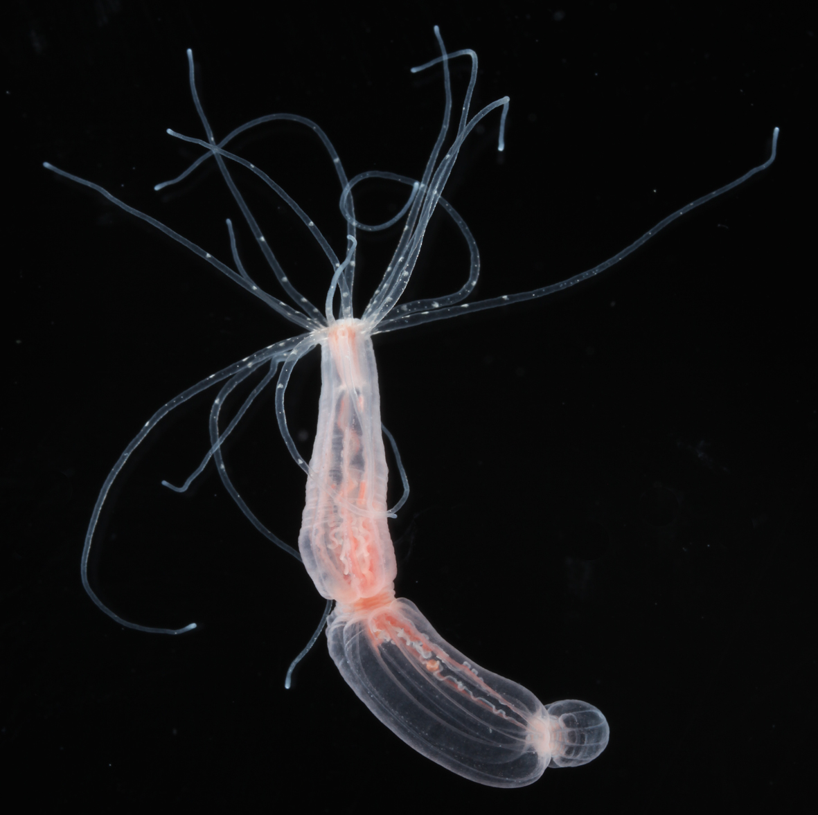
Image of an adult polyp of Nematostella Vectensis.
There are still large knowledge gaps in understanding the molecular mechanisms initiating and implementation of neurogenesis and the regeneration of nerve cells. Being a Cnidarian, Nematostella belongs to the sister taxon of bilaterians, and their nervous system have evolved from the same common ancestor as ours. It is thus of great interest to understand these mechanisms since this could help us better understand how to treat degenerative diseases or injuries affecting the nervous system in us humans.
Ablating and studying the nervous system in Nematostella is made possible by the generation of a transgenic line, Elav1::NTR-Cerulean, with Elav1 being a promoter only initiating
transcription in neurons, NTR a gene encoding for an enzyme called Nitroreductase that can convert a specific antibiotic to a toxic compound, and finally Cerulean that is a probe coupled to the NTR gene, making nerve cells fluorescing.
As a student working in this project, I got to execute my own experiments where I got to ablate the nervous system and then use a confocal microscope to observe the reappearance of neurons. I also did a parallel experiment with the aim to prove the cell-specificity of the Nitroreductase ablation system. By doing these experiments I got to learn how to feed and maintain the animals.
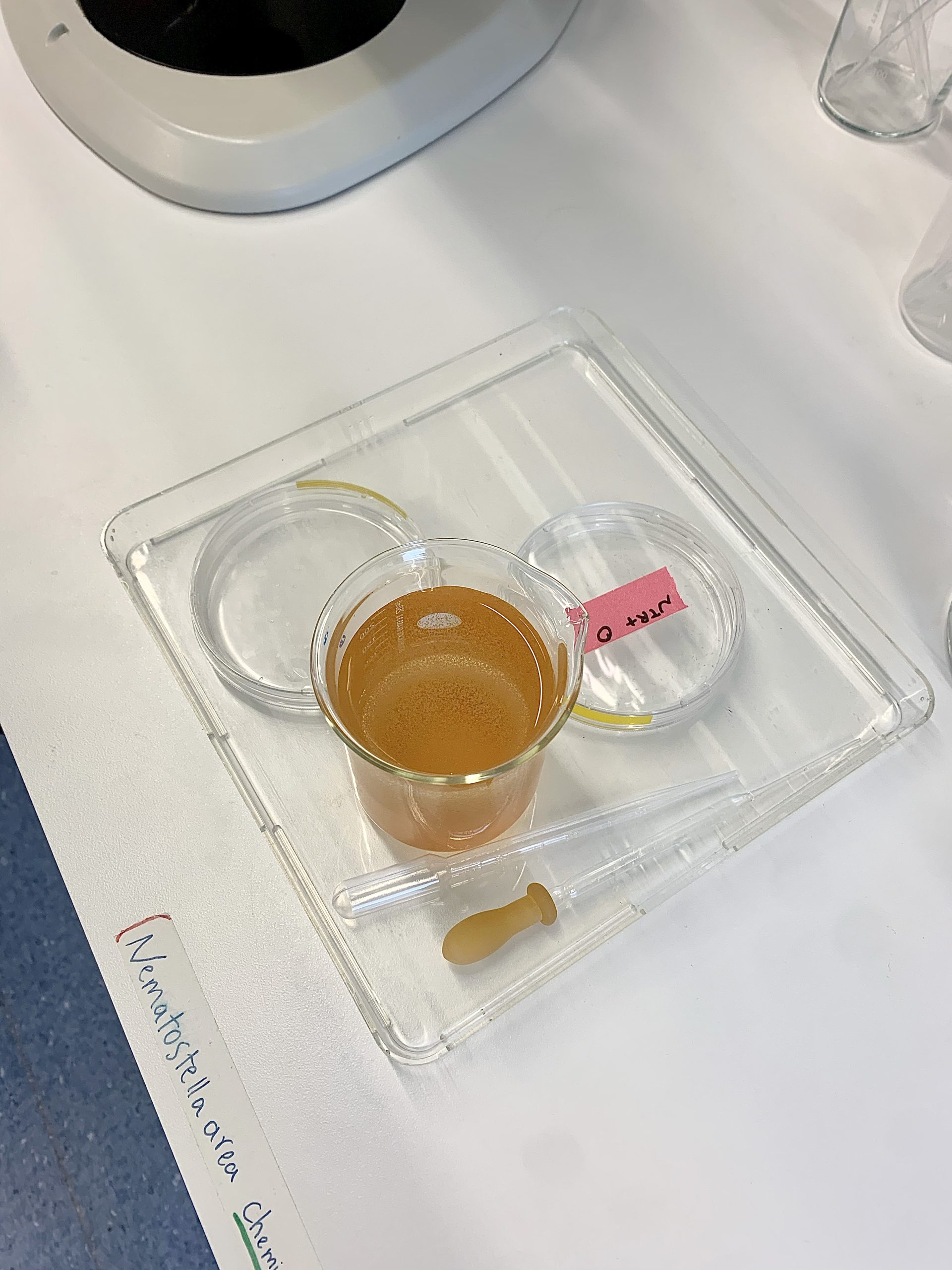
Animals getting fed with Artemia using a pipette.
I also learned how to select the positive transgenic animals using a widefield fluorescent microscope and sort them in different dishes using a pipette. As the positive animals were selected, I got to learn how to prepare the Nifurpirinol (antibiotic) treatment and treat the
animals for 3 days. As the animals were finished with the treatment, I got to prepare the microscope slides and fix the animals on these using a pipette. (This was a lot harder than it sounds). In order to facilitate the imaging, the animals were made paralyzed by adding a chemical called 0.5% Dimethyl sulfoxide (DMSO).
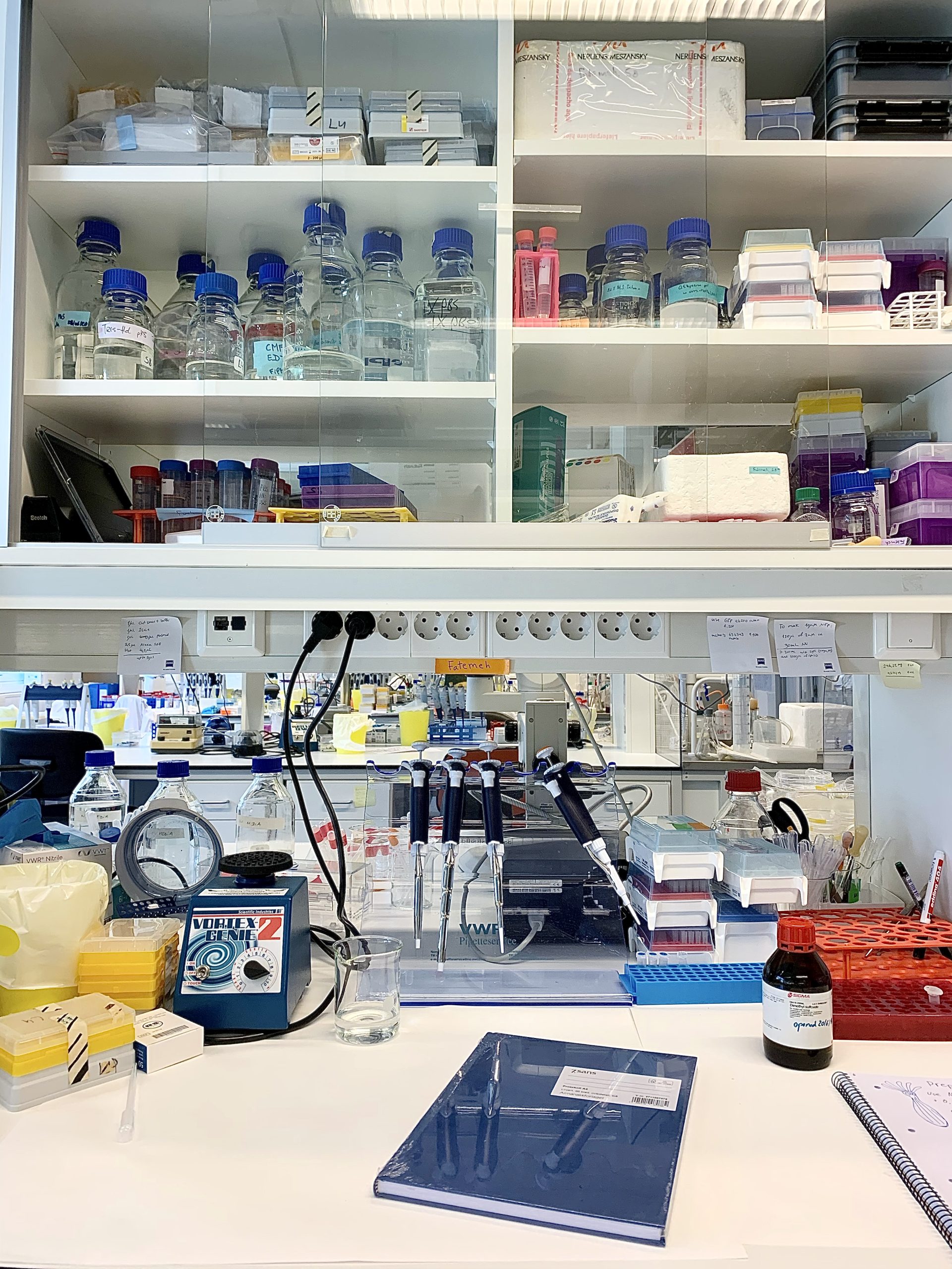
Workspace in the lab where the animals were fed, treated and prepared for imaging.
I was also taught how to use a confocal microscope and as my project preceded, I could do most of the imaging and preparations independently from my supervisor. I really enjoyed that I was trusted to do a lot of the work by myself, and it gave me some confidence. After all the data had been collected – I spent pretty much a full work week only taking images in the
confocal microscope – I got to learn how to use an image program called ImageJ that I would later use to analyse the data and make images for the poster and report.

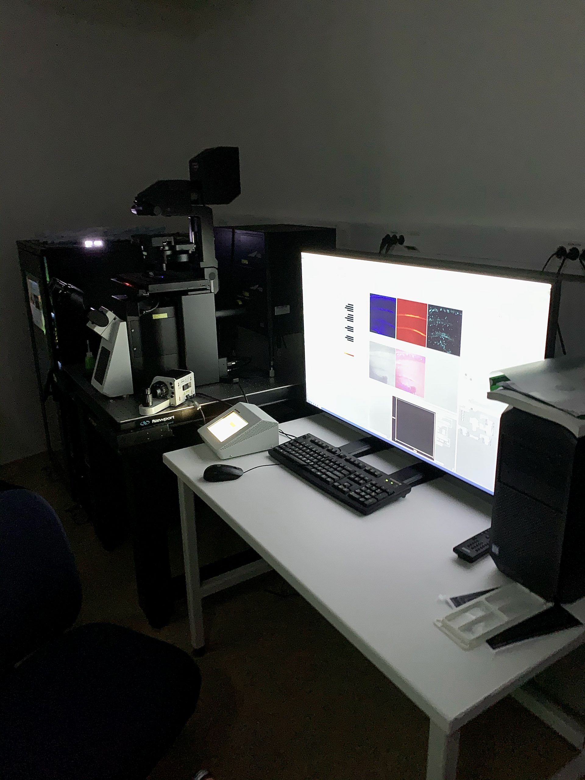
Images from the confocal microscope room.
Learning how to properly use the confocal microscope and the program was definitely no walk in the park but so fun! We had some troubles working out how to properly take the type of images we were interested in but ended up finding another way that worked equally well. Another challenge that arose during this project was that the DMSO made the animals dissolve on the microscope slides as the time went by. This made imaging of the animals a fight against time since we only had so long before the animals dissolved and were not possible to image anymore.
We also had some troubles with some animals dying or getting damaged during the treatment, but overall, the experiments were successful.
During my project we found that cell-specific ablation of nerve cells is possible using a Nitroreductase ablation system. We could also conclude that regeneration of neurons was present at day 4 after complete ablation of the nervous system.
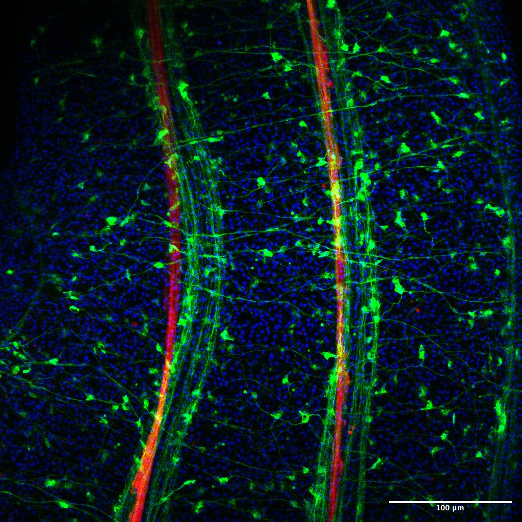
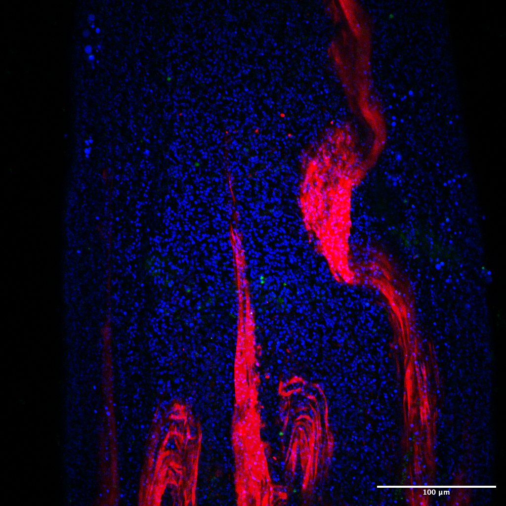
Comparative images taken by the confocal microscope. Untreated animal with intact nervous system to the left and treated animal absent of a nervous system to the right.
The task I enjoyed learning the most during this project was definitely how to use a confocal microscope. I also really enjoyed learning about regeneration from my supervisors and got to take part of paper presentations and discussion with the scientists working in the Rentzsch group.
I would definitely recommend this course to anyone who would be interested in working as a researcher in the future. I feel like I have learned more during this course than most of the theoretical courses I have had during my bachelor, and it is definitely nice with some more practical work as a supplement to all the theory.

Image of fluorescing Nematostella, taken with a confocal microscope.
Thank you for reading!